When Science Is Vital joined with hundreds of other organizations to successfully fend off threatened cuts to the science budget in 2010, the next Spending Review seemed aeons away. By now, of course, most scientists in Britain have heard the persistent rumor that the next Review will be brought forth to some unspecified date in 2013 – in other words, it could be right around the corner. Meanwhile, our economy continues to flounder, punctuated by gloomy headlines predicting that full recovery could be years away. Not, in other words, the climate you really want to be in when the Government starts casting about for further ways to tighten its belt.
This time around, fortunately, we have a lot more than six weeks to make some noise. Science is Vital plans to step into the fray, and we need your help and ideas to put together a new campaign to remind politicians that science is vital for the UK economy.
What can you do?
Come to our first Annual General Meeting, which will be held in Central London on 13 September (7-10 PM, fine details TBA). In addition to the formal AGM, we will have a line-up of interesting speakers and entertainers, plus food and a cash bar, so it should be a fun evening.
To be able to vote, you will need to have become a member within the last calendar year, which you can do by contributing the paltry sum of £3.14 per year (or a multiple of π if you’re feeling generous). Every penny goes towards the business of SIV: we have no salaries or overheads to cover.
Space will be limited, so RSVP as soon as you can.
Our humongous Facebook presence got badly mangled in the platform’s bizarre Group Archiving exercise a few months back, so please go along to our new page and help restore its former glory. And you can support us on twitter by following @scienceisvital.
To convince the Government that science is vital for the UK economy, we’ll need to support the gathering of evidence and behind-the-scenes conversations with politicians. A great way to do this is to join the Campaign for Science and Engineering to enable their superlative work on our behalf. Closer to the time, we should all drop a letter to our MPs and make our convictions known.
Working together, let’s gear up to keep science safe on the other side of the horizon.
The British governmental body responsible for funding research and postgraduate training in engineering and the physical sciences, known as the EPSRC, has been getting some badpress recently. But I couldn’t help being impressed by their new fellowship policy, which was pointed out to me by fellow OT blogger Sylvia McLain.
As my regular readers will know, I strongly object to post-docs being judged against arbitrary sell-by dates, which do not allow any wriggle-room for personal situations. In other words, there should be no magic amount of time beyond which not achieving a permanent research position constitutes failure. A person who takes time out to raise a family, for example, might need a bit more time to publish the same amount of papers as a person who is unencumbered – but this does not mean that the latter deserves an academic research career and the former does not. Rather, let each vie in open competition for the same fellowships and be judged on their own merits. In last year’s Science is Vital careers report, we found that many younger researchers objected to being categorized in this manner, and had been personally affected by its fallout. For this reason, we specifically called for “the abolition of eligibility criteria that effectively discriminate against older postdocs or those who have followed a non-traditional career path.”
Eligibility conditions based on years of post-doctoral experience or permanent academic tenure will no longer apply; as this doesn’t allow for variations of career paths across the EPS disciplines. … A person specification will be used to describe the desired attributes for each career stage, shifting the focus away from eligibility defined by years of post-doctoral experience towards a competency-based approach with an emphasis on the skills and attributes that leaders (and aspiring leaders) need to be able to demonstrate.
I am thrilled to see a research council making a commitment to judging people on their own merits in this way, and I do hope it is the start of a trend that might spread to all the others. Quality comes in many forms, and the most obvious candidates are not automatically the best.
We are usually one step removed from the science that we fund. As taxpayers, we delegate to government bodies the decision about where and how much cash is allocated. Even if we give to specific charities, we can’t control which specific scientists or projects will end up benefiting. On the whole, this is probably sensible: most people don’t have the time, expertise or desire to review all of the thousands of great projects out there and make these agonizing decisions.
But in the brave new age of social media, some scientists are making direct appeals for your personal financial support via dedicated fundraising websites. This idea of boutique science funding was new to me until my good friend Bill Hanage told me about his own activities. Bill is currently an associate professor at Harvard, and is in possession of formal grant funding for his main endeavors. But meanwhile, out back in his sandbox, something very interesting is brewing: a fascinating microbial genomics project he’s running with his intrepid collaborator, Siouxsie Wiles of the University of Auckland, that needs your cash.
We are attempting to track how infectious agents adapt to a new environment, using state-of-the-art imaging and genomics. Siouxsie does the hard graft in her lab, and then sends the DNA to me for the sequencing bit. The costs of genome sequencing are now such that a contribution from one person really can make a difference. And one of the exciting things about this way of doing science is that even if the only thing people reading this do is update Facebook with a link, or tweets it, they are helping to spread the word about the research, and maybe find people who want to get more involved. Thank you. The more genomes we can get, the more power we will have when it comes to the sharp end of the analysis.
It’s a cool project, and you can read more about it here. Whatever the results, we should learn something important, and that will make it a lot easier to apply for more money from conventional funding sources. Serious money. Given the appalling numbers of people who suffer and die from infectious diseases, understanding how they adapt is an essential enterprise.
Bill and Siouxsie have already raised $3960 — with two more days to go. Do consider spreading the word – or even chipping in.
One of the annoying things about getting old is resenting change. So when you’re a scientist, it doesn’t help that the lab environment is one of the most mutable places on earth. New technology emerges all the time, and our brains have to cope — even when ‘improvements’ often seem, to well-greased neural pathways, as unwanted and cumbersome diversions.
Of course some changes are good: I’ve enjoyed swapping X-ray film for a fluorescent image analyzer, and I don’t miss grappling with large amounts of radioactivity one little bit. Cutting and pasting bits of DNA together ye olde fashioned way was certainly therapeutic, but I’m happier with the faster, new-fangled recombination methods that allow you to bypass ligations altogether. No, it’s the useless cosmetic changes that I find annoying: like Microsoft Word deciding to completely rearrange all of its hundreds of functions into entire new categories, so that simple tasks like counting words or changing the font size can take fifteen minutes of hunting through endless tabs and menus. (Dear God, why?, I heard one of my colleagues moaning just the other day as he tried to justify his margins.)
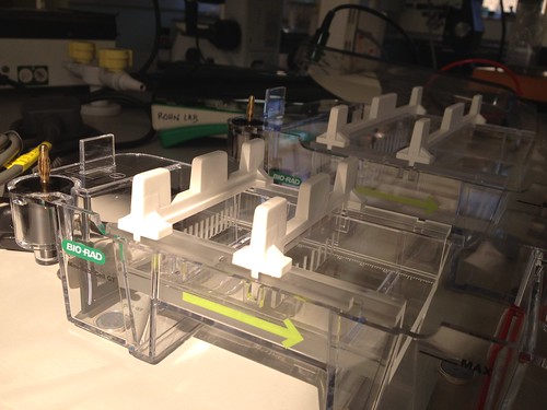
So, grumpy not-quite-as-young-as-I-used-to-be woman that I am, I was really cheesed off when I removed my shiny new electrophoresis boxes from their packing peanut nest and encountered these lime-green arrows on the side:
OK, so it’s a helpful little tip aimed at reminding the uninitiated that DNA is negatively charged and needs to migrate towards the positive pole. I get why they’ve done it. But it just seems… wrong. If you need an arrow to understand which way to load your samples, you probably shouldn’t be trusted with an experiment that mixes liquid and high voltage in the first place.
Plus, the designers who decided to add these helpful arrows probably never worked in a lab where the post-docs routinely swapped the red and black plugs on the power pack to make sure that the PhD students were paying attention.
I’m trundling along here in my new lab, still trying to get everything up and running. On the tissue culture front, things have been fraught for some time, what with delays installing the carbon dioxide and nitrogen tanks, with learning how to live in harmony with the experimental prokaryotes sharing the same air space – all while finishing off a few key applications for funding. Finally, however, I thought I had the system working smoothly, and was ready to perform my first real experiment: infecting bladder epithelial cells with patient bacteria isolated from fresh urine.
This is an absolutely straightforward procedure. I’m simplifying it a little bit, but in essence, you seed your cells onto a coated glass surface (such as the chamber slide pictured above), perform the infection and, some time later, wash the cells and then fix and stain them to inspect what the bacteria have done to the cells and vice versa. It will be my bread-and-butter assay, the basis for testing the genetics and cell biology of infection and also trialling some of the interventions we hope to discover and perfect. So in that sense it is also a bottleneck: the basic experiment without which little else can progress.
So I was pretty miffed when it became apparent that Step One of this simple procedure – the step so basic that you wouldn’t normally even spare it a thought – was just not working. In short, I could not get the bloody cells to stick to the slide. Now this isn’t some ultra-finicking primary cell we’re talking about here: it’s ‘T24’, a full-out transformed tumor cell line that’s been growing for yonks, that’s so happy in culture that you have to split it multiple times per week to keep it under control. These are cells that are obviously quite keen to adhere when the notion suits them. They are the Sultans of Stickiness. They have gorgeous, frilly pseudopods that fan out as they probe their environment and inch their way across the plastic surface, forming and dissolving sticky adhesions with ease as they go. I could gaze at them for hours under the microscope, admiring their luscious lamellipodia.
But on glass, the game was completely off. Many cells can’t grow directly on glass, so when that failed, I tried coating with poly-D-lysine, fibronectin, collagen, serum, and attachment factor. All failed: the cells remained round, clinging together desperately in increasingly large clumps that wafted around the slide like seaweed on the ocean floor before dying a tragic death.
The real mystery, however, began when I contacted the lab in Cardiff that had kindly posted me the cells in the first place. Yes, T24 grew just fine on glass, I was told. No, no coating was required. Yes, they were perfectly happy, in fact, on the precise make of slides that I was using.
So, Dear Readers – I throw the question open. What now? Can anyone help solve the mystery? Or will my line of research suffer…a sticky end?
It is human nature to feel that you’re at the center of the universe, with all of life and experience revolving around your fixed point of view like a lazy orbit of galaxies. On a larger scale, this biased perspective can influence groups of people, and indeed entire countries. Most of you are probably familiar with the light-hearted maps of Daniel K. Wallingford from the 1920s, showing particular cities and their skewed view of the rest of the world, which have been used as inspiration for dozens of similar efforts since that time (The Saul Steinberg cover for the March 29, 1976 issue of The New Yorker being possibly the most well-known example).
Scientific culture, in particular, can be incredibly parochial, especially if you are fortunate enough to do research in a country regarded as ‘world-class’. I have noticed a particular attitude amongst some American scientists, for example, whose words and behaviors can sometimes betray elitism. “Real” science is done in America, you see, whereas other countries are just mucking about and possibly filling in some useful blanks.
A great example of this was the reaction I experienced as PhD student in Seattle, when I was fortunate enough to secure my first post-doctoral position with one of my heroes at the Cancer Research UK London Research Institute. Expecting universal excitement and approval from my professors, I was instead treated in many cases to puzzled looks and bewilderment. I will never forget the words of the most censorious: “It’s all very well and good to do a post-doc abroad, but you do realize it won’t count, and you’ll have to start over with another post-doc in America.”
I don’t know what was more shocking: that this guy would say this to my face, or that he would actually believe it in the first place. (For context, the person in London I was going to be working with was about a hundred times more well-known and famous than this guy. A quick Google search, incidentally, reveals that this censorious professor dropped out of science many years ago, whereas my first post-doctoral supervisor is now a Fellow of the Royal Society. Despite this, the memory of his opinion of the scientific calibre “abroad” still has to power to raise my blood pressure in a swift flash of remembered anger.)
There are of course many other examples of this. You’ll sometimes see American open-access crusaders making – unconscious, I assume – statements implying that all peer-reviewed papers are paid for by American tax payers. We’ve also heard stories about important findings in faraway countries that are not celebrated until a prominent Western lab rediscovers and publishes the same thing in a bigger journal. Even Sarah Palin’s incredulity about “Fruit fly research in Paris, France” is channeling some deeper anxiety about self and other.
But it’s not limited to Americans – I want to stress that this sort of bias occurs in every place, on every scale. When I worked in the Netherlands, my entire universe seemed to shrink to a tiny nation six feet below sea level, and suddenly the lab heads at the Netherlands Cancer institute were perceived to be the top dogs, looming much larger than overseas celebrity scientists, whereas my own situation at the University of Leiden was considerably more lowly. Within Leiden, our biotech environment was looked down upon still further by the surrounding academic labs. Here in London, I sometimes get the feeling that some of my colleagues view the rest of the United Kingdom – aside from Oxford and Cambridge – as being somewhat less lofty. The joke is, of course, that the true beacon is still America, even here: there used to be a joke circulating amongst British PhD students that you really needed a BTA (“Been To America”) on your CV to be taken seriously in your future academic job stakes.
Perhaps the parochialism that most infuriates me is the habit of geographically biased name-checking during scientific seminars. I’m sure you’ve all heard this: Joe Bloggs up on the podium, attributing every background finding to specifically named (typically American or local) lab-heads, until he gets to one particular nugget of information that is attributed to “a Japanese group”. The underlying implication is that this person is no one you need to know about or remember, because he or she performed the research in a faraway place where labs are indistinguishable from one another. But again, it’s all relative. At a recent conference I attended, a British speaker name-checked American and British authors, but referred to “a French group”. A few talks later, a French speaker name-checked French and German lab heads, but dismissed another finding as being from “The Chinese”. And so it goes – each person in his or her own universe, carefully drawing lines between self and other, between tribe and not-tribe, between those belonging in the inner named circle, and those doomed forever to anonymity.
But after knocking around in this new position for nearly three months, I have to admit that it’s rather good fun being poor. When you lack items that you’ve always taken for granted, you have to come up with all sorts of creative workarounds to do your experiments. I’m not exactly fashioning a Large Hadron Collider out of twigs, tin foil and gaffer tape, but until our next big grant gets funded, there are a few things I really need that we just can’t afford to buy outright.
As it’s Friday, I thought I’d share with you a few of our more ingenious inventions:
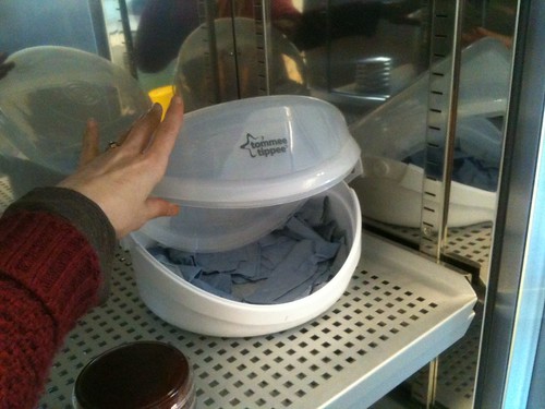
1. The poor man’s CO2 incubator:
As I described last time, we have to culture both sterile human cells, and human cells infected with bacteria. But we’ve got only one CO2 incubator, and the sterile human cells get priority. How then to culture the manky stuff? We can get around the CO2 problem by using media that’s buffered to be at the right pH under normal atmospheric conditions, and we have a regular dry incubator for growing bacteria on agar. What about the requirement for achieving over 90% humidity, though? In hunting around the piles of discarded junk we’ve been slowly getting rid of, I stumbled across a strange plastic container called a “Tommee Tippee”. I had no idea what it was at the time (now I know it’s a baby bottle sterilizer, bought in for someone else’s bizarre experiment a few years back), but without its inset, it looked perfect: a sealable container large enough to contain both a tissue culture plate and a bowl of sterilized water – all nestled on moist paper towels.
It worked a treat! All our cells were still alive and happy the next morning. Sorted.
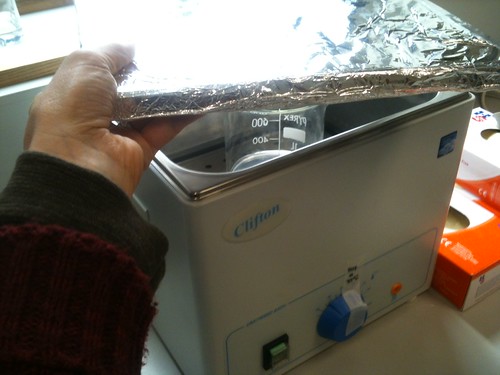
When I arrived at the lab, I was bequeathed a pile of nearly-new stuff bought off a previous researcher’s grant – the woman had moved on to a non-research job. One of these was a water bath – without a lid. Had it been lost? When I went on the manufacturer’s website to see if I could order a replacement, I realized immediately why the previous researcher had not ordered one: it was sold separately, and it was nearly the same price as the bath itself. After another rummage in the Pile O’ JunkTM, I came up with an appropriately sized piece of plywood; after a nice coat of aluminum foil, it was good to go.
Result: a bit more prone to fungal infections than a real lid, and liable to tear, so we have to change the foil once a week. Still, preferable to sacrificing £150 quid!
Price: Hardware: FREE. Consumables (foil, to be replaced at weekly intervals): Approx 5p/week.
3. The poor man’s N2 tank (a cautionary tale):
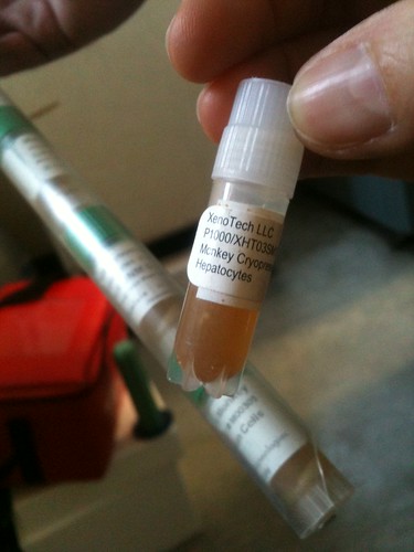
OK, so liquid nitrogen is pretty damned cold: −321 °F, to be precise. You can’t just store your samples in tupperware or a Thermos flask. We had no recourse but to buy a proper 30 liter tank, but we went the second-hand route. This wasn’t easy: the UK is a very small country, and there is hardly anything out there. However, we eventually struck gold, purchasing our tank from a charming Yorkshireman called Steve, who was even nice enough to throw in a three-month guarantee, and a random set of storage canes for free.
All fine and dandy, except it soon became apparent that Steve hadn’t bothered to look inside the used tank before he shipped it off:
But no problem: after throwing away the ancient samples of Funky Monkey, we were ready to place a fill order with our friendly neighborhood BOC rep.
Price: Hardware: £400 + VAT (retail price, about £3,500). Consumables (nitrogen): Approx £6/week plus delivery. Free vials of dead monkey hepatocytes: Priceless.
4. The poor man’s shaking bacterial incubator:
Although originally a microbiology lab, the room and kit I inherited had no provision for growing broth cultures of bacteria. Traditionally, you need an incubator with a mechanism for shaking flasks and tubes with extreme vigor so that your bad-assed bugs are properly oxygenated and give high yields. This one was a real bitch to work out, but with a bit of trial and error, my colleague and I cobbled together the following little beaut of a solution.
1. Take tiny 37-degree benchtop incubator and put it on its side on top of a magnetic stir plate.
2. Open the small metal hatch (of completely unknown function) that is now on the ‘floor’ of the incubator, in contact with the stir plate.
3. Sterilize a 25 mL Erlemeyer flask with a magnetic flea inside it.
4. Add water for test run; pierce foil with thermometer to monitor internal temperature.
5. Place flask inside hole left by open hatch (almost a perfect fit, coincidentally) and switch on magnetic stirrer.
6. Turn on incubator and tweak the unmarked temperature dial until a stable 37°C is achieved.
Hypothesis: a vigorous flea-stir will create sufficient oxygenation for the bad-assed bug cultures.
Result: Unknown as of yet – still waiting for my consignment of tryptone and yeast to arrive. But we did manage to keep the test water temperature between 36 and 38°C over a weekend. Unknown as yet whether the incubator will work reliably long-term upended precariously on its side.
It was always going to be a difficult relationship.
We knew from the very start that they weren’t very well-suited. After all, they came from such different backgrounds. They were used to such radically different environments. They scarcely even spoke the same language, and they certainly held radically disparate views about the best way to lead their lives. Despite everything, we held hope in our hearts that somehow, they’d manage to set aside their differences and co-exist in harmony.
Alas, we were wrong.
Yes, phase one of an important sociological experiment in now completed in my lab, rather like a reality TV show featuring the world’s most incompatible roommates. And I’m sad to report that it’s Eubacteria, 1…
…and Eukaryotes, nil.
So what do you do when you suddenly find yourself in charge of a lab that needs to culture both sterile human cells, but also bacterially infected human cells? And you only have one tissue culture room?
The room in question had been the domain of microbiologists when I arrived. Nobody was doing regular cell culture. There were two sterile hoods, both of which were used to culture infected patient cells – and for some reason, despite the presence of seven entire empty bays in the main lab, all nine undergraduate students were also using the same small room to streak out all their bacteria onto agar plates. The place was practically humming with prokaryotic vibes.
So I banished the students, the Petri dishes and the bacterial incubator, ordered the room and its hoods stripped down and organized a deep clean of every conceivable surface with detergent and bleach. After a week-long quarantine in which no one was allowed in, I imported a clean CO2 incubator, CO2 tanks, a water bath and a new fridge. The right-hand hood was to be for sterile tissue culture only, and the left was for experiments with bacteria in them – only when sterility was absolutely necessary. No reagents were to be shared between the two users. In addition, no bacterially infected cultures were allowed in the new gas incubator; instead I bought in some CO2-independent media and rigged up a moist chamber inside the dry 37-degree incubators for the microbiologists to use (using an old baby-bottle sterilizing chamber we found up on a shelf – don’t ask). Then and only then did I order my bladder epithelial cells, thaw them out and put them into culture.
There were a few unavoidable shared elements allied to the two separate workstations. We have only one centrifuge, so while I bleached a dedicated set of adapters for cell work, there was no avoiding the fact that the two camps’ samples would have to share the same air space while spinning. There is only one inverted microscope, too, so sterile and infected plates sit for a time on the same stage. And of course, the air inside the TC room might be wafting a few hundred CFUs more than the normal atmosphere at any given time. Nevertheless, I thought it we were all careful and considerate, it just might work.
I cultured my cells happily for a week with no problem – and then the first microbiologist came in to do an experiment in the left-hand hood. A particularly manky set of samples, I noted uneasily – so full of rods that the medium was cloudy. The day after, on a hot afternoon when the temperature probably hit 30 degrees inside the sunny room, I taught one of my colleagues how to split cells and charged him with maintaining the line from then on. Just before the Easter break, one flask was contaminated, and today, the other one succumbed as well. I don’t know if it was caused by the incompatible roommate problem, the heat, or simple newbie bad luck – after all, many beginners get contaminations in their first month at it. Too many variables to say for sure.
We’ll wipe everything down and try again. What else can we do?
I’ve just written to my MP, Simon Hughes (Liberal Democrat, Bermondsey and Old Southwark), airing my feelings about the Government’s hasty and ill-advised bill to track email, web and Skype communications of any citizen without just cause. A copy of my letter is included below. If you live in the UK and have strong feelings too, you can write your own MP via They Work For You.
Dear Mr Hughes,
I am writing to you to express my fervent opposition to proposed Government plans to allow the Government Communications Headquarters to monitor the email and web communication of ordinary citizens, without a warrant. This a shocking breach of privacy – and even if it advances our security interests by a few degrees, it surely cannot be worth the cost to our dignity as a civilized society.
I can only echo what Davis Davis wrote in the Sun on this issue, as quoted by the BBC news website: “We already have a law which lets the secret services eavesdrop on suspected criminals and terrorists. The new law does not focus on terrorists or criminals. It would instead allow civil servants to monitor every innocent, ordinary person in Britain, and all without a warrant. If they want to see all this information they should be willing to put their case before a judge or magistrate. This will force them to focus on the real terrorists rather than turning Britain into a nation of suspects.”
I do hope I can count on your support.
Yours sincerely,
Dr Jennifer Rohn
Principle Research Associate, Department of Medicine, University College London
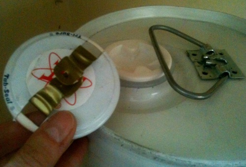
Following on from a discussion about light bulb changing jokes, I was bustling around my lab this morning, getting ready to cryogenically preserve some backups of my new bladder epithelial cell line. I was all set: I’d ordered the Mr Frosty container, prepared my freezing medium, and the five plates of cells were looking beautiful under the microscope. Earlier, I’d been assured that I’d find isopropanol in the flammables cupboard.
The isopropanol was there, all right: a giant, Fort Knox of a metal canister that had never been opened. And it didn’t look like it wanted to be opened any time soon. I’ve never seen anything like it before, so I googled the two registered trademarks printed on the opening: Tri-Sure and Tab-Seal. This led me to a 2-page pdf instruction manual (and a few YouTube videos) on how to open the canister – with a dedicated tool called a “Tri-Sure universal plug wrench”.
Having just spent a month clearing out this lab, I couldn’t recall having unearthed anything that looked quite like said wrench. Neither could one of my old-timer colleagues, who I’d brought upstairs to poke and prod at the impregnable tin.
At this anxious juncture, we finally noticed the two unassuming workmen who’d been lurking in the corridor between the main lab space and some of its annexes, climbing up and down ladders and whistling quietly to themselves. They were, as it happened, changing lightbulbs – a few of the fluorescent strip lights, and the dodgy emergency exit sign. Noticing the impressive array of tools in his box, we asked one of them for help. He didn’t have a Tri-Sure universal plug wrench, and he politely brushed off my attempts to show him the instruction manual on my mobile phone – but nevertheless he radiated confidence. Sure enough, in about thirty seconds, after employing a screw-driver as a wedge and a spanner as a grip, the canister was open.
So though we still don’t know how many scientists it takes to change a light bulb, I now feel sufficiently clued in as to how many light bulb changers it takes to break into a Tab-Seal closure and save the day. Which really just goes to show that sometimes, a PhD is not all it’s cracked up to be.
By day: cell biologist at UCL. By night: novelist, broadcaster, science writer, sci-lit-art pundit, blogger and Editor of LabLit.com. I blog about my life in science, not the facts and figures.